The fundamentals of x-ray technology have barely changed in decades. But the systems surrounding it are constantly shifting.
Grant Smith, CRES, knows imaging. As assistant director of clinical engineering at Duke University Health System, Durham, NC, Smith helps to manage a professional team that includes 15 imaging engineers. Before Duke, he was an in-house imaging specialist himself, with West Penn Alleghany Health System. And prior to that, beginning back in the early ’90s, he spent his workdays with a third-party service organization—again, tending to the needs of a variety of imaging equipment.
And yet, Smith says, despite that experience, when it comes to imaging, and the equipment surrounding x-rays in particular, he continues to learn new skills all the time. “It’s such a rapidly changing market that a lot of the things that I used to be really good with are no longer in service,” he says. “And the digital environment—it’s completely different. You’re no longer dealing with electronics that are specific to devices. Everything now is controlled by PC, and that computer basically runs the system for you.” It’s been a shift, Smith says, that has forced him and his colleagues at Duke not only to become fluent in the “physics and safety requirements around the technology,” but also to shore up their IT capabilities—and in many cases, to hire people who already have them.
In fact, Smith says, his hiring tactics are a major part of his overall approach to x-ray and imaging system management and maintenance. “We always have crosshairs on individuals with specific skillsets,” he says. “Especially people who understand the higher-end modalities, like CT and MR and linear accelerators. It’s really difficult to bring somebody in with no experience and train them in this environment.”
For those who work on the x-ray side of things (magnetic resonance imaging and ultrasound, for example, do not rely on x-ray components to acquire images), “a minimum requirement even to be considered would be an associate’s degree in electronics technology or the equivalent,” Smith says. Most of his hires have taken courses in radiography and fluoroscopy technology and connectivity, and most receive further training—through courses led by manufacturers themselves—on the exact devices the facility keeps on its floors.
“If we’ve purchased Philips digital x-ray systems, and that’s what we’re using, they need to know how to work on Philips digital x-ray systems.” Smith likens their recruiting of imaging experts to the way a Toyota dealership might look for a mechanic: They’d want to hire people who had experience with Camrys, he says, “because that’s what they’d see day in and day out.”
“Stable and Steady”
Interestingly, while the equipment that relies on x-ray technology for its imaging is in the midst of the same digital revolution as every other medical device, x-ray technology itself has hardly budged in decades. “The actual development of the x-ray hasn’t changed,” Smith says. “We’ve seen some interesting changes on the CT side, in its use of an x-ray tube and how that works—but really, the same thing always happens.”
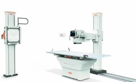
That process is familiar to most biomeds. Each x-ray machine includes an electrode pair. Inside the machine, a high-voltage current passes through a filament (the cathode), heating it up and releasing electrons. Those electrons are drawn at high speed across a vacuum (the x-ray tube), where they collide with the anode material, typically a heavy metal like tungsten. This collision generates energy as x-ray radiation, which passes through the patient’s body to generate a pattern that can be recorded as an image.
When it comes to x-ray generation, agrees Mike Harvey, founder, partner, and vice president at Mikron Digital Imaging and director of business development with Meridian Medical Systems, Aurora, Ohio, the technology behind the process is about “as long-lasting, stable, and steady” as it gets. “It’s basically just a power supply and an x-ray tube,” he notes. “And it’s been that way forever.”
Actually, Harvey says, the last major advancement in x-ray technology took place in the 1980s with the move to high-frequency generators—and that, he says, means that healthcare facilities (and biomed departments) that wish to cut costs should look to imaging. “Instead of buying a new integrated digital radiography system,” he says, where the digital components are deeply entwined with the x-ray generator, “we think it makes more sense from a financial standpoint to buy the very solid, reliable, and unchanging analog x-ray system separately” from the DR system. (His company makes an ultra-high-definition DR unit called the Meridian Monarch that operates off a PC tablet.) “That way,” Harvey says, “you can move from DR system to DR system as that technology evolves.”
The financial advantage of doing it this way is clear, Harvey adds, since an integrated DR system might cost $250,000 or more, while the DR components on their own run closer to $60,000 to $70,000. “When you buy an integrated system,” he says, “after 5 years it’s like owning an iPhone 3.” The system might work, that is, but it will probably seem inadequate compared to its competitors.
In practice, most facilities upgrade their digital radiography technology when their physicians demand that they do so, Harvey notes. “Really, they’re the ones who draw the line. They see that the standard of practice in DR imaging has changed, and they look at their device, and if it’s not keeping up with what their peers are using, it’s going to seem deficient.”
The operative word here is “seems,” Harvey adds. “In fact, it may still make perfectly good images. Film made perfectly good images. DR computer radiography made really good images. But it’s the software, and the image processing. It’s the ease of use. When a product reaches a certain age, you start to run into problems with performance that can’t be solved on the platform it’s on.”
Meridian itself has run into this issue with the software that accompanies its products. “For the longest time our software operated on a 32-bit Windows XP platform, and then the 64-bit platform became available and we were stuck with a program that was slow compared to the newer stuff,” Harvey says. Eventually, the company “hit that tipping point where we decided we needed to change,” so they migrated their old platform into a 64-bit format and changed the specifications for the device they were selling it on.
“The day eventually comes where you ask yourself, ‘OK, even though this system still makes images, does it do it as effectively and efficiently as this other system does?’ ” If the answer is “no,” Harvey says, and if making the switch will save time, money, or both, “then it’s time to make the move.”
On the Floor
Maintaining efficiency while minimizing expenditures is a top priority at any facility, of course, and Duke University Health System is no exception. To keep things moving in Smith’s department, his imaging experts are divided into six groups that only work in specific areas: ultrasound, MR, CT, cardiac catheterization and interventional, radiology, and radiation and oncology. This way, he explains, his engineering staff can spend their days focused on the imaging technologies they’re most familiar with, and not have to learn every last detail about every device in the facility. Preventive maintenance (PM) of the various devices takes up a significant portion of their time, Smith says, but PM demands typically pale in comparison to the hours they devote to service events: over the course of one recent month, his team responded to nearly 300 service calls and did 102 PMs.
When it comes to x-ray equipment, Smith says, the most common issues seen by his team concern mobile digital detectors, especially their handling. “We have everything from digital portables to integrated rad rooms, and when they get dropped, that’s a huge liability for us to manage.” There’s the expense, he says, but there’s also the fact that there’s often nothing the technician can do—or at least not before speaking with the manufacturer. “These things have so few parts, and you’re usually dealing with a visual substrate. They’re not easy to work with.”
Duke’s solution, Smith says, is in the parts-and-service agreements it arranges with its vendors. “It’s just like having car insurance. The plans themselves are expensive”—typically $7,000 to $20,000—“but they’re nowhere near the price you’d pay to buy a new device.” As it stands, when a digital detector breaks, “it’s almost always either exchanged or replaced,” he says. (Meridian encourages its customers to take the same approach: Its top-of-the-line DR detector is paired with a Panasonic 20-inch 4K tablet that comes with a 3-year factory warranty, and the Meridian also offers a no-fault drop-protection program for both the computer and the detector. “If you drop it and it breaks,” Harvey says, “we ship you a new one.”)
Service and Support Options
The service needs of bigger and less fragile imaging devices are a different story. For them, Smith notes, his trained technicians can often handle the most difficult problems right there on the hospital floor. Still, most facilities must at some point call for outside assistance with their imaging technology. When that happens, they typically do one of two things: contact a third-party service company like Technical Prospects in Appleton, Wis, where Jeremy Probst is the chief operating officer, or go directly to the manufacturer.
Technical Prospects, Probst explains, only deals in Siemens equipment, and provides parts, training, and support for everything Siemens makes. Students in one of their courses, he says, might spend two full weeks in lectures and labs getting in-depth instruction on how to maintain and fix Siemens CT scanners and x-ray systems, for example. The courses offer a “hands-on experience,” Probst says, and covers topics including preventive maintenance, troubleshooting of problems, replacement of parts, and calibration procedures.
A lot of facilities are “trying to bring their imaging work in-house and get away from the OEM,” or original equipment manufacturer, says Technical Prospects’ director of sales, John Vandersteen. “They might come to us because the manufacturer has told them they’re not going to support a certain piece of equipment, or that they need to upgrade or buy something new because that product is at end of life,” when in fact that device may be perfectly safe and functional. “We might be able to support that equipment for another 10 or 15 years by recycling parts,” Vandersteen says. For all those facilities where money is tight, “that’s an easy way to keep costs down.”
Another option: forge a relationship with a major manufacturer in the style of the “Managed Services Alliance” between Philips Healthcare and Georgia Regents Health System (GRHealth) in Augusta, Ga. Announced in the summer of 2013, the $300 million agreement calls for Philips to provide GRHealth with a comprehensive range of technologies and services at a fixed monthly cost over 15 years. And according to Philips’ Kelley Connolly, senior director, Managed Services Operations and Implementation, the maintenance and upkeep of the health system’s imaging equipment is included in the contract. Like many institutions across the country, GRHealth had been operating with a fleet of very old analog x-ray equipment: “It worked, so they kept using it, and they’d had it for 15 or 20 years,” Connolly says.
By 2014—just 1 year into the agreement—Philips had replaced the organization’s 15 C-arms and 12 mobile x-ray systems with digital fleets, Connolly says. The company also installed a new picture archiving and communication system to create a “single, integrated clinical IT platform” that gives clinicians in any department across the system easy access to patients’ imaging records.
The digital upgrade transformed GRHealth’s overall workflow, Connolly says. “When we stepped back and asked, ‘How does mobile x-ray fit into the overall enterprise concerns of the hospital?,’ it really had to do with patient discharge. So when stat orders come through in the morning, and you need to get patients out the door by 11, having digital x-ray [radiographs] that can be transmitted wirelessly to radiology to read—that really speeds things up.”
Another advantage in digital mobile x-ray systems: They can be used in the emergency department. “It’s about not having to transport patients down to radiology,” Connolly says. “And not only do we just bring a mobile [unit], but then the mobile doesn’t have to wheel all the way back. So you’ve eliminated half the transport requirements.” In the grand scheme of things, Connolly says, “X-ray service is not a big worry in most hospitals. But in the context of length of stay, overall patient throughput, and driving out cost with vendors, there’s a lot of opportunity.”
The Future of X-Ray
In the future, says Duke’s Smith, the fundamental technology behind x-rays is likely to remain the same as it’s been for the last three decades. What will change, he predicts, is the vehicle by which those images are displayed. “Instead of those panels that are so clumsy and delicate and so expensive to insure, we’ll probably start to see some really cool, flexible devices where it would be absolutely fine if you dropped them.” He pictures a device that looks a lot like the cheap, roll-up, plastic sleds that were popular in the 1980s. “Look at the smartphone today versus what it was 10 years ago. Or TVs today compared to what they used to look like. Everything is getting thinner and smaller. Technology is evolving, and I can only guess that we’ll see a lot of the same themes” in the hospital environment.
Harvey of Meridian Medical Systems agrees. “Sometime between now and the time where images just appear in the air, the hardware—the detector itself—is going to look like a sheet of paper,” he predicts. X-ray technology has evolved from fixed detectors, which were built into walls; to tethered detectors, which could be moved as far as their cords allowed; to wireless detectors, which could be relocated as long as they could still communicate with the system built into the room. And now, Harvey says, “We have wireless detectors that can communicate with a tablet and run around the entire hospital.”
X-ray technology is x-ray technology, he notes, and it’s highly unlikely it will ever change. But the devices that make x-rays useful, and the skills that are needed to keep those devices running? “That’s where it gets interesting,” he says.
Chris Hayhurst is a contributing writer for 24×7. For more information, contact editorial director John Bethune at [email protected].
Lead photo: © Leaf | Dreamstime.com