The medical community has focused for many years on encouraging every woman of the appropriate age to participate in a Screening Mammography Program. Its goal is early diagnosis of breast cancer, although the recommended age for screening varies depending on which organization or group you talk to. We are not here to debate the merits of early detection or how low a lifetime dose should be in order to be safe.
Our goal here is to explore the various imaging technologies that are used to enhance the presentation of breast tissue when either the breast is too dense for traditional screening methods or an evaluation of tissue invasiveness is required. Other uses for some of these adjunct technologies are to determine the effectiveness of treatment for symptomatic tissue and for treatment planning and post-treatment surveillance.
Traditional Screening Mammography
The basic mammography screening series is a four-image set consisting of craniocaudal and mediolateral oblique images of each breast. Screening mammography is approximately 97% effective for the estimated 60% of women with normal breasts (those with about a 50/50 ratio of glandular to fatty tissue.)
For the approximately 40% of women who have what are considered dense breasts, screening mammography is less beneficial. In fact, screening effectiveness can be as low as 48% for dense breasts. This group includes young women whose family histories indicate they should undergo earlier than normal mammograms. It would also include women with implants, large breasts, or those with higher percentages of glandular tissue than others in their age groups.
Low energy x-ray is the primary method of acquiring mammographic screening images. Over the years, technology has progressed from basic film to very specialized films to full-field digital images. Current digital imaging technology now includes tomosynthesis, which is a synthesized 3D image produced by acquiring multiple images from various angles and processing them in much the same way that computed tomography does. While tomosynthesis shows much promise, as of this writing it has yet to be recognized as a standard of care.
Ultrasound’s Role in Screening
Originally intended as an adjunct to traditional screening mammography, ultrasound has increasingly been used as a follow-up imaging procedure to further analyze findings or to better demonstrate difficult-to-view anatomy from screening images.
For dense breasts, manually scanned ultrasound images have been used to provide image data to supplement the screening process. More recent developments in ultrasound such as automated breast ultrasound (ABUS) have elevated ultrasound to an approved imaging technique for screening of asymptomatic women with dense breasts by the Food and Drug Administration (FDA). GE ABUS was the first FDA-approved ultrasound screening mammography system, with others sure to follow. Some other names for similar technology are automated breast volume scanner (ABVS) and automated whole breast ultrasound (AWBUS).
Adjunct Imaging and Clinical Images
Clinical images are directed by findings during the screening process. Depending on the nature of the anomaly, additional images may be ordered to further diagnose the suspected symptomatic observations. These images can be either additional mammographic images using different angles, spot compression, and magnification, or manually guided ultrasound imaging.
Adjunct or supplemental imaging modalities are employed to assist in achieving an accurate diagnosis when traditional mammographic studies don’t demonstrate tissues correctly or to better determine the extent or status of a lesion.
For symptomatic women, supplemental imaging can be used to evaluate the invasiveness of cancers, therapy planning and to evaluate changes in masses during treatment. Once treatment is completed, it may seem intuitive that one of these supplemental imaging techniques would be used for ongoing monitoring. However, in most cases basic screening mammography would be indicated.
Let’s take a look at some of the supplemental imaging technologies used for breast imaging and discuss where they excel and what their limitations may be.
Ultrasound
Ultrasound is a very versatile imaging modality. By using different modes and system configurations or advanced software processing, clinicians can realize a wide array of benefits.
Manual ultrasound exams are primarily used to evaluate tissues identified through screening. Operated in brightness mode (B-mode), this modality is particularly good at differentiating cysts from solid masses. That strength is due to ultrasound’s ability to observe the difference in the speed of sound in liquid and solid mediums.
Automated Breast Ultrasound (ABUS) uses a large linear probe with a slight curve that moves in a straight single sweep across the breast. The large curved compression paddle achieves a more uniform compression over the entire breast. The curvature in the probe results in the convergence of the scan line energy and thus greater penetration. The shape also enables better detail resolution at depth.
Automated Whole Breast Ultrasound (AWBUS) uses a smaller linear probe that is automatically guided through an overlapping pattern to acquire the entire field in multiple passes. Once these individual images are stitched together, they create diagnostic images ideal for evaluating large areas to help locate satellite masses.
Water Bath Ultrasound (WBUS) requires the patient to lie facedown on a table with her breast hanging pendulous into a vat of water. The image is acquired from around the breast, and image presentation is similar to that of a CT scan. The water provides a very good acoustic coupling, which makes WBUS suitable for dense breasts.
3D Ultrasound Imaging generated by ABUS, AWBUS, and WBUS allows for improved visualization of breast structures and is essential for thorough evaluation of dense breasts. 3D imaging is also valuable for what is referred to as staging. Staging shows the relationship between satellite masses and a dominant tumor.
Tissue Harmonic Imaging is preferred for determining lesion conspicuity and for margin analysis. The process takes advantage of a couple different principles. First, fluids generate a weak harmonic signal when compared to soft tissue. This difference makes evaluation of complex cysts easier, because cysts are commonly filled with fluid. Second, fatty tissues have a stronger harmonic signal than soft tissue, which makes solid masses stand out from the surrounding tissue.
Doppler Ultrasound uses the shift in high-frequency signals to assess blood flow throughout the body and can therefore prove useful in evaluating breast tumors. Vascularity assessment differentiates one class of tumor from another. Central, penetrating, branching, and disordered blood vessels are signs of a malignant tumor; therefore, malignant tumors tend to show color flow signals at a higher rate than benign tumors. Likewise, hypervascular breast cancers tend to be of a higher histological grade and are considered more malignant.
Computed Tomography
Standard CT images, acquired by scanning axially around the body of the patient, can be used to image breast tissue. The subject lies supine on the table, which prevents the breast tissue from pulling away from the chest wall. This posture makes it more difficult to isolate specific breast tissue along the chest than tissue closer to the nipple. CT imaging of the chest region requires a large field of view, reducing the CT system’s ability to resolve very small structures. This modality is therefore better suited for large mass evaluation than for imaging micro calcifications.
Breast CT imaging systems specifically designed for mammographic imaging use cone beam CT technology. These systems have a smaller bore where the breast hangs down pendulous, while the x-ray tube and a large field detector rotate around it. This design allows the field of view to be smaller, which improves resolution over standard CT, but the modality still has some limitations resolving small micro calcifications. Where breast CT excels is in its ability to detect and visualize breast anatomy and soft tissue masses. In fact, breast CT outperforms conventional mammography in this area.
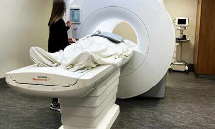
Magnetic Resonance Imaging
Breast MRI claims an extremely high sensitivity and the ability to differentiate between malignant and benign masses. Breast MRI is used in conjunction with, but not as a replacement for, conventional imaging. Even given its advantages, MRI is too time-consuming and expensive to be a practical replacement for standard x-ray.
Annual MRI Screening is recommended for the following groups:
- Patients with BRCA gene mutation. A BRCA mutation is a mutation in either the BRCA1 or BRCA2 gene. Harmful mutations in these tumor suppressor genes produce a hereditary breast-ovarian cancer syndrome in affected families.
- Patients who are untested for the BRCA mutation but have a BRCA-positive first-degree relative.
- Patients who received radiation to the chest between the ages of 10 and 30 years old.
- Patients who have a lifetime risk of greater than 20% to 25% for the development of breast cancer.
Undergoing treatment for breast cancer is not itself an automatic qualifier for annual MRI screening; once treatment is completed, mammograms and ultrasound are primarily recommended.
Postlumpectomy Breast MRI is also useful as a follow-up imaging modality to look for residual disease after a lumpectomy has been performed.
Nuclear/Molecular Imaging
Nuclear or molecular imaging is very good at demonstrating tissue functionality in terms of metabolic activity. A higher metabolic activity indicates a more aggressive cancer. This imaging modality, despite having a high rate of detection and specificity for malignancies, is not recognized as a high-resolution imaging modality.
Nuclear Imaging involves administering a radiotracer isotope by injection. The radiotracer typically combines a chemical that would naturally migrate to the site of a mass and a short half-life radioactive isotope such as Technetium-99. Angiogenesis (the formation of blood vessels) surrounding the tumor allows for increased delivery of the radiotracer and causes more radiation to collect in the mass. The gamma radiation emitted by the isotope is observed by scintillating materials in a flat detector and conversion to an electrical signal to be processed into an image. Depending on the number of detector heads, the image can be either two-dimensional or three-dimensional.
Positron Emission Tomography (PET) is very similar to conventional nuclear imaging in its use of radiotracers. However, because the detectors are located all around the patient, PET produces 3D images of the radiotracer concentrations. Using biochemicals like fludeoxyglucose F18, PET can be helpful in determining the possibility of cancer metastasis, or the spreading of cancer to other sites in the body.
PET/CT pairs the ability of PET imaging to accurately present the metabolic activity of tissues with the anatomic resolution capabilities of CT, and combines both individually acquired scans into one image.
Dual Head Molecular Imaging, because of its specialized detectors, has a resolution capacity far superior to that of its predecessors. In fact, its resolving capabilities can be comparable to those of conventional mammography. Radiotracers are still required to produce the image.
Thermal Imaging
Thermal Imaging systems observe the temperature of tissues and present images that are color mapped to indicate the variances in those temperatures. Typically, higher temperatures are shown in red while cooler temperatures appear blue. Thermal imaging relies on the principle that more metabolic activity and vascularity cause more heat.
Maintaining Today’s Women’s Health Department
All of this technology associated with women’s health departments will ensure a place in the industry for service professionals who can master the skills required to maintain it.
The variety and complexity of these imaging modalities will require teams of professionals to maintain them. The vast majority of service professionals working in women’s health will find themselves servicing the digital mammography systems and ultrasound units that are used for screening and clinical imaging, which do the bulk of the work. Fulfilling a general imaging service role, they may not recognize how important they are to the effective delivery of women’s health services.
Dale Cover is president and chief operating officer of RSTI. For more information, contact chief editor Jenny Lower at [email protected].
Photo credit: © Stratum | Dreamstime.com