Expert insights about one of imaging’s buzziest modalities
If the buzz at the recent annual meeting of the Radiological Society of North America (RSNA) is any indication of market trends, the ultrasound sector is hotter than ever. In this expert roundtable, four industry insiders—Dan Skyba, director, Ultrasound Business Unit, Canon Medical Systems USA, Inc; Neal Sandy, chief marketing and commercial officer, GE Healthcare Clinical Care Solutions; Mike Justice, senior territory manager at Trisonics Inc; and Amanda DePalma, head of global marketing for ultrasound at Philips—sit down with 24×7 Magazine to discuss some of the biggest innovations showcased at RSNA 2018 and what healthcare professionals should know before purchasing ultrasound equipment.
24×7 Magazine: How has the ultrasound market evolved in recent years? How do you expect it to evolve even more in the future?
Dan Skyba: The ultrasound market has been growing at a tremendous pace, with projections going up by the hundreds of millions each year. This growth is due to a few important factors that have converged in the ultrasound market. Ultrasound systems are now on the market for every clinical use, at every price point, and they all deliver high-quality imaging. Riding on the tails of the exponential growth in computing power and the miniaturization of hardware, ultrasound has benefitted tremendously. Over the past 20 years, there has been a dramatic improvement in image quality, resulting in ultrasound images that often rival those of CT, and even MR. Speed and ease of use has improved significantly and, at the same time, the costs have decreased.
Ultrasound will continue to grow rapidly and will outpace every other imaging modality. The decreased size and cost of ultrasound units, combined with machine learning and automation, will simultaneously add value to today’s users, as well as bring ultrasound to completely new markets. [Moreover,] we’re seeing ultrasound make the move from a diagnostic tool to a screening tool. And continued improvement in image quality, automation, ease of use, and faster connectivity will bring ultrasound to new environments outside of the hospital or clinic and, hence, expand the broader global marketplace.
Mike Justice: The ultrasound market is evolving exponentially. The U.S. market should see around 5% financial growth this year, with Asia overtaking it in market growth for the first time. I am seeing more interest in the specialty market such as musculoskeletal (MSK), critical care, ER, and vascular. In the past, point-of-care systems only provided a basic set of controls and had poor image quality. Those days are long gone, however.
Today, point-of-care systems are marketed to specific departments and have a wide range of options that used to be exclusive to premium console systems. I expect that, soon, the typical premium console system will look a lot like the point-of-care system that we see today. There will be no compromise of image quality with portability.
Amanda DePalma: We continue to see strong growth in the ultrasound market, [due to] affordability, access, and safe technology that helps individuals across the care continuum, which give us the ability to reach a more diverse patient population and continue to improve millions of lives around the world. We also continue to see advancements in automation, quantification, and the use of anatomical intelligence to make ultrasound a more objective and intelligent solution for both patients and providers.
This year, Philips launched Lumify with Reacts, an integrated tele-ultrasound solution that integrates live communications support into the ultrasound exam for a more meaningful collaboration between care providers both inside and outside the hospital setting. Ultrasound will continue to become more accessible, in the coming years.
Neal Sandy: Ultrasound users and uses continue to expand. For example, we’ve witnessed a proliferation of ultrasound systems in many new clinics within hospitals. As more applications of ultrasound emerge, new clinical personnel gain interest. This also has and will continue to influence the need to specialize ultrasound devices for specific applications and workflows.
24×7: Manufacturers always showcase their “latest and greatest” innovations at RSNA’s annual meeting. What were some of the top advancements in ultrasound showcased at RSNA 2018, which took place in late November?
Sandy: Artificial intelligence (AI) was front and center for ultrasound at RSNA. GE Healthcare, for instance, showcased several commercially available AI applications on the Logiq E10, Voluson E10, and Venue systems. These AI solutions are deployed to support applications for lesion detection, OB measurements, vascular flow, fetal heart, fetal brain, and diagnosis of shock. We also highlighted our most advanced cardiovascular system, the Vivid E95. Additionally, the Vscan Extend had new onboard clinical support applications including LVivo EF—the first auto-ejection fraction function on a handheld system leveraging AI technology licensed from DiA Imaging Analysis.
DePalma: One of the clinical areas Philips is making advancements in is breast exams. At this year’s RSNA meeting, we showcased our new ‘ultimate ultrasound solution for breast assessment,’ available on both the Philips EPIQ and Affiniti ultrasound systems. This integrated breast solution combines the eL18-4 transducer, anatomical intelligence for breast, ElastQ imaging shear wave, and new precision biopsy for comprehensive screening and diagnosis. With this new breast [technology], clinicians will be able to efficiently assess, monitor, and treat breast diseases, increasing diagnostic confidence and helping to improve patient care.
Skyba: This year, Canon Medical Systems introduced the industry’s first 33 MHz ultra-high-frequency linear iDMS transducer, available on the Aplio i800. This transducer uses iDMS technology and a single-crystal-wide band to provide superb resolution and detail in ultrasound imaging.
We also announced the addition of our Contrast Vector Imaging (CVI) tool to our Aplio i800 to provide additional information on liver hemodynamics and expand the use of contrast-enhanced ultrasound on this ultrasound system. The CVI tool helps visualize vascular structure inside lesions by tracking individual contrast bubbles and analyzing the direction and velocity of each bubble to help support a more confident diagnosis. This new tool has the potential to help clinicians better see complex lesion vascular networks—seeing clear delineation of feeding vessels with quantifiable perfusion and direction information.
24×7: Which clinical area(s) do you expect to see ultrasound moving into most? And what technological advancements have been made to ultrasound transducers used in this specialty?
DePalma: We expect to continue to see advancements across all clinical segments. We know clinicians need better solutions that not only improve detection and diagnosis of disease, but also increase throughput and productivity while maintaining the highest levels of confidence. At Philips, we are addressing today’s clinical challenges by providing comprehensive solutions across key applications like liver, small parts, and breast. By harnessing the power of advanced technologies with clinically tailored tools, clinicians have the ultimate ultrasound solutions to help provide patients with the diagnosis and treatment they need.
Justice: I believe the high-frequency technology market will see the most growth. The MSK and breast imaging market seem very strong. I was recently involved with an installation of a system designed specifically for the critical-care market. This system is an example of a specialty system designed for one department only. It has a very large touch screen monitor, over four-hour battery life, and specific software applications for the critical-care doctor. The manufacturers see that there is a need for specific point-of-care applications, and they are starting to deliver.
As for technological advancements, current trends seem to be based on efficiency and patient throughput. The applications are providing more and more automation of the image quality and calculations, which reduces the patient scan time. The transducers are also starting to be part of this concept. Some systems have buttons on the transducer that are programmable—a new feature that allows the transducer to be selected by double-tapping the head with your finger—and new array technology that is increasing the image quality and bandwidth.
Sandy: We see an expansion into more acute care applications, as well as into general practice physicians and family medicine. From a technology perspective, ultrasound transducers are getting smaller, lighter-weight, automatic, wireless, and better integrated with sophisticated software image-forming technology. We’ve also seen a step function change in computing power with graphical processing units, which allows a pivotal shift in the acquisition of the ultrasound beam. This change offers a new way of processing ultrasound and eliminates the need for traditional controls to provide consistently improved image quality from the skin line all the way down deep into the image.
Skyba: We’re seeing the biggest innovations happen around imaging of the liver. As the obesity epidemic grows globally, nonalcoholic steatohepatitis, nonalcoholic fatty liver disease, and other diseases of the liver are on the rise. Ultrasound is a great tool to help distinguish between different disease states in the liver and can help clinicians determine the best treatment plan. New liver analysis tools—based on shear-wave technologies and contrast—have been a game-changer for measuring tissue characteristics in the liver.
Ultrasound tools that quantify liver tissue viscosity, attenuation, and perfusion patterns are available and all contribute important diagnostic information. At Canon Medical, we offer a Liver Analysis Package on our Aplio i-series platform, which is designed to gives clinicians the tools they need to address every stage of liver disease.
Another area of innovation is in matrix technology transducers. 3D ultrasound is still a hot area, but now matrix technologies are being applied to 2D probes to create images of superb quality. Technologies such as dynamic micro-slicing (iDMS) create beams which are thinner to greatly improve spatial resolution. Ultra-high frequency probes are also pushing the boundaries of clinical imaging. Canon, for instance, now has transducers at 24MHz and 33MHz, which can resolve structures at the 100-micron level.
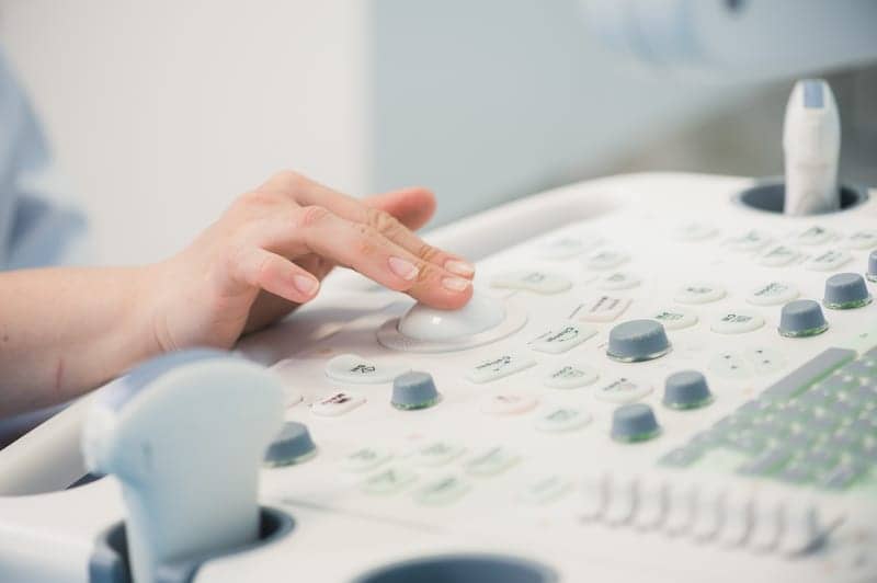
24×7: What best practices should HTM professionals adhere to when servicing, maintaining, handling, and repairing ultrasound devices—and ultrasound probes, in particular?
Justice: The most important practice is to prepare for a complete loss of data. This would include a backup of all configuration and preset files, all license options, and making sure that you have the current software on hand, including any patches that were released. [You should also] make sure that the departments are completing a configuration backup after they make major changes to their system settings. Protocol settings can take hours to build and will be lost with a failure and software reload.
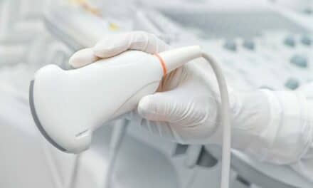
Another best practice is to test transducers for leakage in a water bath, while taking precautions to ensure the electrical safety of the system. There can be microscopic holes in the lens that are not visible to the human eye and will leak excessive amounts of current to the patient.
Probes are very important to maintain properly. They are expensive, fragile, and the point of contact to the patient. Always do a complete visual inspection of the probe, from connector to the lens. Look for cracks in the case, holes or delamination of the lens, and any damage to the cable or cable strain relief. Remember: Most transducers can be repaired if you discover the damage at the early stages. Trisonics, for instance, has received probes that were damaged beyond repair simply because the end user continued to use the probe with a damaged strain relief. The damaged strain relief was allowing gel to be pushed in to the case, eventually making it to the array.
Skyba: Care should always be taken when transporting equipment and maintaining probes according to the manufacturer’s recommendations. But more recently, the medical community has been challenged with a greater threat to their equipment—that of malicious cyber-attacks, which can cripple not only a unit, but an entire hospital network. The recent Wanna-Cry virus is a perfect example.
The good news is that Canon ultrasound has put in a layered approach to equipment security that keeps the system safe from attack. Anti-virus, whitelist programs, and data protection are available on all systems. [Specifically,] Canon ultrasound follows the stringent Risk Management Framework put in place by the Department of Defense, and was recently certified with Authorization to Operate to be installed at the most sensitive military and government hospitals.
Sandy: Ultrasound probes are complex medical devices that should be handled, stored, cleaned, and serviced per the guidance of the original equipment manufacturer. Probes are critical components of the ultrasound device, particularly given that they come in contact with the patient. To ensure high image quality and patient safety, probes must be cleaned and reprocessed per the manufacturer’s guidance.
24×7: What special features should healthcare facilities look for when purchasing ultrasound equipment?
Sandy: Our customers like their ultrasound equipment to be specific to the clinical applications most needed by their department. One size does not fit all, as workflows, clinical applications, probes, and digital needs vary depending on the use case. Because of this, we offer a broad portfolio and work closely with global clinical experts to develop our equipment by care area. Customers should also consider whether a company’s equipment is designed to advance and expand over time as new technologies emerge. Understanding the lifecycle of equipment is imperative in making an overall good selection for the hospital or clinic’s needs.
Justice: This is a tough question because most special features are specific to different types of scans and modalities. For instance, one feature that is important to the MSK market may not apply to the cardiology market. I think the most important practice to use when purchasing is to make sure that all the options that the end user needs are turned on. Most special features are sold as software options and are enabled through keys. As an example, I have been called out to network a system that the customer purchased from another vendor and found that the DICOM feature was not enabled. On the quote, make sure that you request which features are enabled on the system—and then verify [their existence] when you take delivery.
Skyba: When purchasing ultrasound equipment, healthcare facilities should look for a partner with equipment that helps deliver clinical precision, departmental productivity, the highest level of cybersecurity, and features that support sonographer health. Recent studies have shown that 90% of sonographers are scanning in pain. Canon’s Aplio i-series platform, for instance, includes a feature called iSense design to make our systems easy to maneuver.
Specifically, with an over 36 cm panel height adjustment, a lateral slide, and a fully articulating monitor arm, providers can optimally adjust the console to virtually any scanning position. We also recently partnered with Sound Ergonomics to cultivate a new program called “Healthy Sonographer,” which aims to help prevent sonographer injuries through education and best practices sharing. As ultrasound expands to new territories, these considerations will remain critical in helping customers deliver diagnostic imaging that offers clear images for confident diagnoses—and supports the overall health of the industry.


I like what Justice said about how healthcare facilities need to look for features that are specific to their market, since markets like MSK and cardiology will have different required features. When I think about it, the many advancements in ultrasound technology have probably led to a lot of unique options for the machines. Thanks for sharing this interesting info about ultrasound systems!