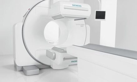
Coming soon to a radiology department near you is a new star attraction: the PET/CT hybrid. If perchance this exciting performer has already debuted at your local theater of operation, then you have no doubt thrilled to all that PET/CT maintenance offers. If not, then please sit back and enjoy the following preview, which has been approved for all audiences.
PET/CT is a whole-body imaging modality that physically mates a positron emission tomography (PET) scanner to a computed tomography (CT) machine for the most comprehensive views possible of tumors, cardiac health, brain functionality, and more. Useful in both clinical and research applications, PET/CT reveals the location of those problems radiologists suspect are present and need to be found. As icing on the cake, it also shows the degree of metabolic activity present there.
 |
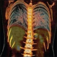
“PET by itself is like looking at a globe with all the topographical features, while CT adds the lines to show the borders of all the countries—in other words, when you put PET and CT together, you get a more complete picture of what the body looks like inside,” says John Nieberle, assistant product leader for service with GE in Waukesha, Wis. His colleague, online technical support engineer Steve Surprenant, offers this explanation of the technology: “CT gives you the physical makeup of the internal structures, and PET gives you the functional part of that same anatomy. So, if you’re looking for possible tumors inside a structure, PET/CT will show you not only the suspected site, but it also shows you what that growth is doing.”
The patient who undergoes a PET/CT scan begins the process by being placed on a table that feeds into the gantry of the CT machine. The system then generates a CT image of that patient. When the image-making data are all collected, the table then advances into the PET scanner’s gantry where a PET image is produced. Upon completion of that process, a computer takes the output from each component modality and merges the data into a unified image.
PET/CT users also can choose to make images with one or the other of the two halves. For example, some facilities utilize it for CT diagnostic work when its other CT scanners are busy, according to Ziad Abumughaiseeb, a customer service engineer in San Francisco who works for Siemens Medical Solutions.
Well-Built Systems
The uniting of the two modalities in this manner introduces all the maintenance and service requirements familiar to PET and to CT individually, plus offers a few additional new ones.
Bob Pray, CRES, senior field service specialist with CHI clinical engineering at Porter Hospital, Denver, says that the current generation of PET/CT scanners are well built and reliable. That, he contends, means clinical/biomedical engineering departments can expect to encounter little in the way of big problems. “Of the two machines, the PET is the one that can be expected to have the fewest serious problems because it has fewer moving parts than the CT,” Pray says.
Conversely, the most commonly encountered minor problems seem to reside with the PET component, reports John Klink, BSEET, imaging services specialist, Memorial Medical Center, part of the Sutter Health system, Modesto, Calif. “The problem we see more of than anything else on the PET side is occasional drift of one or more of its roughly three-dozen detector modules,” Klink says. Prior to joining Memorial about a year ago to work in-house full-time, Klink worked for 23 years as a field services engineer for GE Healthcare. “When drift occurs in a PET detector, the remedy is to recalibrate the affected module or modules. That’s a process that takes about 30 minutes.”
And 30 minutes is about all the time a hospital can afford to lose—longer than that and the morning’s delivery of the nuclear tracer isotope used for the PET portion of PET/CT scanning can spoil, Klink warns. The isotope is fluorodeoxyglucose, abbreviated as FDG or spelled in proper long form as 2-fluoro-2-deoxy-D-glucose, if chemistry is your passion. It is expensive and has a radioactive half-life of just under 2 hours. In the case of Memorial Medical Center, FDG is produced at a cyclotron facility about 1 hour away in the city of Sacramento. The isotope product is out the door for the rush trip to Memorial almost the moment it is packaged. “The concern with the detector recalibration time constraint is not just about money lost in the event the FDG is wasted,” Klink mentions. “Machine downtime also means studies are not being performed, and that in turn leads to lost revenue.”
Another commonly encountered minor bugaboo with PET/CT is slowed image processing. This, according to Pray, is usually due to memory overload in the system’s computer. “It normally takes about 10 minutes for a PET/CT computer to process the captured image data,” he says. “When the memory on these units starts to become full, the processing speed drops. To speed it back up, you have to periodically delete the oldest studies held in memory. You should do this on a daily or weekly basis, but you know it’s definitely time to perform this cleanup if the radiology technologists inform you that the system is taking 30 minutes or longer to process a study.”
However, slow processing also can be the fault of traffic on the network that links the PET/CT with the radiology department’s PACS informatics system. Says Pray, “Sometimes you’ll have other modalities attempting to send studies to PACS at the same instant the PET/CT is.” The biomed is responsible for telling the difference between a network problem and one that originates with the PET/CT scanner’s memory, he reminds.
What Can Go Wrong?
Among the problems you hope never occur with a PET/CT is jamming of the calibration tool. The reason is that the tool features radioactive components of 1.5 millicurie (5 millicurie on older versions). The radioactive parts stay within a shielded housing until needed for calibration. Then, they are retrieved for insertion into the scanner by a mechanical arm. “If something hangs up and causes a failure of the housing door to open or close, you’ve got to manually resolve the problem,” Pray says. “What’s dangerous about this is you’re dealing with a mega-source of live radiation. You have to suit up in protective clothing and be assisted by a physicist with a Geiger counter.”
Another truly nasty problem is arcing of the CT slip rings. Fortunately, this seldom happens, Klink assures. “The slip rings are important because all communication between the rotating frame and the stationary frame of the PET/CT system pass through these,” he says. “The slip rings are in mechanical contact 100% of the time with power brushes and signal brushes. The PET gantry is behind the CT gantry, and the slip rings are behind that. As a result, access to the CT gantry to service or replace the slip rings requires detachment of the PET scanner.”
Naturally, detachment is an involved task. So is reattaching them, because alignment of the two is critical. “We’re talking tolerances of only a few millimeters, and if your adjustments are outside those specs, the consequence is going to be misalignment of the PET image and the CT image in the composite image produced by the system software,” Klink says. “If that happens, the doctors, when they look at the image, will be led to believe the cancer is in this location here when it’s actually in that other one over there.”
Scrupulous attention to preventive maintenance (PM) can help avert both of these loathsome problems.
PM Protocol
Early on, Siemens developed a PM protocol that serves as something of a gold standard for the care and feeding of PET/CT. This protocol calls for a quarterly PM, beginning with a visual inspection of the equipment. During this step, the hardware is eyed for signs of physical damage and the surrounding environment is assessed for conditions that could harm the machinery, according to Abumughaiseeb. Another step entails checking that the electrical voltage and current powering the system are within acceptable ranges. Later in the process, the brushes and fluids associated with the system’s major moving parts are checked.
Whatever set of PM protocols you follow, experts recommend keeping on hand a stock of lower-cost but higher-usage replacement items such as belts and filters. “The basics,” Surprenant says. And, as with any mission-critical modality, PET/CT maintenance and servicing should be scheduled for those times when shutting down the system will have the least impact on daily clinical operations. “A typical PET/CT inspection takes 4 to 6 hours to complete,” Abumughaiseeb says. “Some biomeds like to spread those hours over a couple of days to leave themselves time to work on other projects or urgent tasks.”
Abumughaiseeb believes that most PET/CT PM work can be handled by one biomed. The remainder—such as replacement of certain components in the gantry—require a second set of hands. No special tools should be necessary for conducting a PM, although Abumughaiseeb suggests that carrying the tools offered by Siemens for recalibration and other odds-and-ends tasks is a good, step-saving idea. “Always be prepared and ready to go,” he offers.
Meanwhile, PM at Porter Hospital for the PET/CT is incorporated into daily rounding. “I give our system a 15- to 20-minute inspection each morning to make sure there weren’t any overnight problems with it,” Pray says. “I time my visit to coincide with the firing up of the system so that I can see whether it comes up correctly. I also spend some of that time asking the technologists if they’ve noticed anything out of the ordinary with the system.”
Eyes and Ears
Some hospitals fully assign the task of PET/CT PM and all other service requirements to the equipment manufacturer, while others split the service duties between their in-house team and the OEM. Biomeds, meanwhile, need to remember that their role with PET/CT includes serving as the eyes and ears of the OEM’s field engineering team and, as such, they must be continually prepared to provide good technical information on demand to facilitate faster service action.
One important bit of technical data they should have handy for the second-call squad is the system’s error logs and any other recorded messages, Surprenant suggests. However, if a remote connection has been established between the hospital site and the vendor, those event logs can likely be uploaded from the PET/CT system to the online engineer’s computer terminal. “That capability also allows us to run certain diagnostic tests remotely to enable us to work with the in-house team to get the system going again, or to support our field team in their efforts on-site,” Surprenant says.
Second call comes, of course, when the problem encountered exceeds the knowledge and skills of those performing first call. Pray says he strives to push back that boundary by producing detailed incident reports at the end of every OEM-conducted PET/CT service call, adding that he likes to sit in on the repair sessions and take notes. “The way I write these reports is almost like a dissertation,” Pray says. “I do that so that at whatever point in the future this same problem occurs, I can go back and see a thorough discussion of what was done to identify the problem and the steps that needed to be taken to correct it. This is an exercise that helps me become more knowledgeable about PET/CT, and become a more valuable resource for my hospital.”
Rich Smith is a contributing writer for 24×7. For more information, contact .