
 Over the years, microscopes have become an indispensable tool in helping healthcare providers in the diagnostic and therapeutic arenas. Microscopes used in diagnostic applications are generally found in the laboratory area of the hospital. Those used for therapeutic purposes are typically located in the operating room as a tool for assisting with surgery. In this article we will briefly look at each type of microscope and highlight some differences and similarities.
The Basics
To begin this discussion, let’s review some basic information about optics. Wikipedia defines light as a radiant energy found in the electromagnetic spectrum. Light visible to humans can have a wavelength of 380 to 800 nm. However, many sources will refer to the visible light spectrum as being in the 500 nm range with a frequency of 600 GHz.
Some of the primary properties of light are intensity, propagation direction, frequency or wavelength spectrum, and polarization. The speed of light in a vacuum is 299,792,458 meters per second, and is one of the fundamental constants of nature. When light rays hit a glass lens, they are bent according to Snell’s law, which is stated as n sin ? = n’ sin ?’. This explains how light is bent through a lens, whether it be a concave or a convex shape. This bending of the light is referred to as refraction. The principles of optics also apply to other medical devices that may use mirrors in the optics, but they will typically manipulate reflection instead of diffusion. (For a nonmedical but well-known example of how reflection can be used, consider the Hubble space telescope, which employs mirrors and the principles of reflection to focus images.)

Figure 1: Light rays are bent by a lens, creating a focal spot. (Click to enlarge.)
The focal length of the microscope is a measurement of how far it takes the light to be focused to one point, or the focal point. The shorter the distance in which the light rays converge, the higher the magnification factor. This bending or converging the light rays is dependent on the curvature of the lens used. Figure 1 shows how light rays are bent by the lens and the resultant focal spot which is created.
This focal spot is created the same way in either type of microscope and the shorter the focal spot the more magnification is achieved.
Diagnostic Microscopes
A standard microscope in the lab is referred to as a compound microscope. These units will typically have a light source to illuminate the sample slide. This light travels through the iris diaphragm into the slide, which rests on the slide stage, and then up to the objective lens and beyond to the eyepiece. Magnification is accomplished not only in the objective lens, but also inthe eyepiece. Typical eyepiece magnification factors may be 10X, while the objective lens may have typical magnification factors from 4X or 10X to 40X, and possibly as high as 100X.
Magnification factors for a compound microscope can be found to be in the 200,000 range. Once 200,000 magnification has been reached, the human eye cannot visualize the object of interest. Magnification factors are found in the same way as the gain of a Darlington pair of transistors in electronics. The magnification level of the compound microscope is found by multiplying the magnification of the eyepiece by the objective lens magnification factor. So, with an eye piece of 10X and an objective lens magnification factor of 40X, the unit would display at 400 times larger than the object. This amount of magnification will allow visualization of bacteria, molecules, and many other animal cells. However, viruses, atoms, and many molecules are such small particles that an electron microscope is used instead.
Surgical Microscopes
Surgical microscopes differ from lab types in that they usually have much less magnification than standard compound microscopes. The image is also steerable by the surgeon foot controls, so that the surgeon always has use of both hands. Many surgical scopes have a standard magnification on the eyepiece (depending on the type of surgery). The focal length of the microscope is altered by changing the lenses on the bottom of the unit, much like changing the objective lens in a compound microscope.
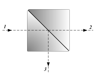
Figure 2: A diagram of a beam splitter. Light comes into the unit (1) and is split into the image the the surgeon sees (2) and the image displayed on a video screen (3). (Click to enlarge.)
High end surgical microscopes may have multiple viewing heads or eyepieces to allow other members of the surgical team to view the surgical field. Some of the more expensive high-end scopes, such as those with multiple viewing stations, may also have video hook ups to allow viewing of the surgical procedure on a monitor and for recording the procedure. Units with video outputs will employ beam splitters to divert a portion of the image to a charge-coupled device (CCD) to form the video displayed on the monitor. The beam splitter is an optical device that splits a beam of light in two different beams for multiple viewing. This device is a crucial part of many instruments such as interferometers, technologies found in radiology, surgical microscopes and other devices. Figure 2 shows a beam splitter.
Surgical microscopes can be used in many types of surgery, but are heavily used in endodontic, ENT, ophthalmic, vascular, and neurosurgical procedures.
I hope you find this article informative and useful once you sit down to your certification exam. Until next time, keep studying!
Review Questions
Answers below
1) The property of bending light with a lens is described by which law?
a) Lenz’s law
b) Snell’s law
c) Ohm’s law
d) Newton’s law
2) Some studies have found the human eye visible light spectrum to be in this range of the electromagnetic spectrum:
a) 20-20000 Hz
b) 300-600 nm
c) 380-800 nm
d) 680-1200 nm
3) To find the magnification factor of a compound microscope, which formula should be used?
a) n sin ? = n’ sin ?’
b) eye piece lens magnification / objective lens magnification
c) eye piece magnification + objective lens magnification
d) eye piece lens magnification X objective lens magnification
4) Which focal spot distance will display the most magnification?
a) 3 meters
b) 6 in.
c) 8 in.
d) 16.24 cm
John Noblitt, MAEd, CBET, is the BMET program director at Caldwell Community College and Technical Institute, Hudson, NC. For more information, contact editorial director John Bethune at [email protected].
Answers: 1—B, 2—C, 3—D, 4—B